Pill Camera
Information
Capsule Endoscopy Is A Procedure
That Uses A Tiny Wireless Camera To Take Pictures Of Digestive Tract. The
Capsule Endoscopy Camera Sits Inside A Vitamin-Sized Capsule You Swallow. As
The Capsule Travels Through Your Digestive Tract, The Camera Takes Thousands Of
Pictures That Are Transmitted To A Recorder You Wear On Belt Around Your Waist.
Capsule Endoscopy Helps Doctors
See Inside Your Small Intestine – An Area That Isn’t Easily Reached With More
Traditional Endoscopy Procedures. Traditional Endoscopy Involves Passing A Long
Flexible Tube Equipped With A Video Camera Down Your Throat Or Through Your
Rectum. However This Traditional Type Of Endoscopy Can’t Visualise The Majority Of The
Middle Portion Of Small Intestine. Therefore Capsule Endoscopy Is Used To
Examine Parts Of The Gastrointestinal Tract (Gi Tract) That Cannot Be Seen By
Other Endoscopic.
Diseases
That Can Be Detected By Capsule Endoscopy
1. Inflammatory Bowel Disease (Crohn’s Disease)
2. Polyps
3. Ulcers
4. Tumours
Manufacturers
This Technology Was Originally
Developed By Gabi-Iddan And Paul Swain With The First Pill
Swallowed In 1997.
What To Expect During A Capsule
Endoscopy
1. 8 Sensors Placed
In On Abdomen
2. Sensor Belt
Strapped Around Waist Over Shirt
3. Swallow Pill
Containing A Camera
4. Pill-Cam
Transmits Images Of Gi Tract To Sensor
5. Disposable
Pill-Cam Will Pass Via Bowel Movement
Side Effects
1. Developing A
Fever After Swallowing The Capsule
2. Having Trouble
Swallowing
3. Beginning To
Vomit
4. Increasing Chest
Or Abdominal Pain
Total Artificial Heart (Tah)
Information
An Artificial Heart Is A
Prosthetic Device That Is Implanted Into The Body To Replace The Biological
Heart. Artificial Heart A Pumping Mechanism That Duplicates The Rate, Output
And Blood Pressure Of The Natural Heart; It May Replace The Function Of A Part
Or All Of The Heart
History
Year
|
Scientist
|
Event
|
|
1953 |
Dr. John Gibbon |
A Heart – Lung Machine |
|
1964 |
The National Heart , Lung And Blood Institute |
Set A Goal To Design Tah By 1970 |
|
1966 |
Dr. Michael Debakey |
Implantation Of A Partial Artificial Heart |
|
1967 |
Dr. Christian Barnard |
Human Heart Transplant |
|
1969 |
Dr. Denton Cooley |
Total Artificial Heart |
|
1982-85 |
Dr. William Devries |
Jarvik Total Artificial Heart |
|
1994 |
Food And Drug Administration |
Approval The Left Ventricular Assist Device |
|
2000 |
Texas Heart Institute |
Jarvik 2000 |
|
2001 |
Abiomed Inc |
Abiocor |
Total
Artificial Heart Prototypes
1. Polvad
2. Phoenix – 7
3. Abiomed Abiocor
4. Syncardia
5. Magscrew
6. Cleveland Heart
7. Abiomed Abiocor Ii
8. Carmat Bioprosthetic Heart
9. Frazier – Cohn
10. Soft Artificial Heart
How Does It Work?
The Tah Replaces The Lower
Chambers Of The Heart, Called Ventricles.
Tubes Connect The Tah To A Power Source That Is Outside The Body. The Tah Then
Pumps Blood Through The Heart’s Major Artery To The Lungs And The Rest Of The Body.
The Tah Has Four Mechanical
Valves That Work Like The Heart’s Own Valves To Control Blood Flow. These Valves Connect The Tah
To You Heart’s Upper Chambers, Called The Atrium,
And To The Major Arteries, The Pulmonary Artery, And The Aorta. Once The Tah Is
Connected, It Duplicates The Action Of A Normal Heart, Providing Mechanical
Circulatory Support And Restoring Normal Blood Flow Through The Body. The Tah
Is Powered And Controlled By A Bedside Console For Patients In The Hospital.
After They Leave The Hospital, People With A Tah Use A Portable Control Device
That Fits In Shoulder Bag Or Backpack And Weighs About 14 Pounds. It Can Be
Recharged At Home Or In The Car.
Good News For Patients
O The Average Time On Support For Syncardia Tah
Patient Is Approximately 130 Days, But The Tah Has Supported Patients For Much
Longer Periods Of Time. In Fact, Several Patients Have Been Supported For More
Than 4.5 Years.
O Stable Tah Patients Are Able To Leave The Hospital
And Enjoy Active Lives At Home While They Wait For A Donor Heart.
O The Tah Is Available At More Than 140 Hospitals In
Over 20 Countries.
Tony Stark – The Iron Man Is The Most Popular Example Of Artificial Heart!!!
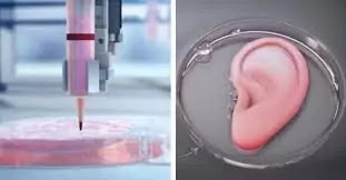
3D Printing Of Organs
Information
3-D Bio-Printing Is A Form Of
Additive Manufacturing That Uses Cells And Other Bio Compatible Materials As “Inks”, Also Known As Boinks, To Print
Living Structures Layer By Layer Which Mimic The Behaviour Of Natural Living
Systems.
Bio-Printed Structures, Such As
An Organ On A Chip, Can Be Used To Study Functions Of A Human Body Outside The
Body (In Vitro), In 3-D. The Geometry Of 3-D Bio-Printed Structure
Is More Similar To That Of A Naturally Occurring Biological System Than An In
Vitro Study Performed In 2-D, And Can Be More Biologically Relevant.
It’s Used Most Commonly In The Fields Of Tissue Engineering And
Bio-Engineering, And Material Science. 3-D Bio-Printing Is Also Increasingly
Used For Medical Applications In Clinical Settings – 3-D Printed Skin Grafts, Bone
Grafts , Implants, Biomedical Devices And Even Full 3-D Printed
Organs Are All Active Topics Of Bio-Printing Research.
The
Types Of Printers And Processes
1. Inkjet Printer
2. Multi-Nozzle
3. Hybrid Printer
4. Electors Pinning
5. Drop-On-Demand
Different Techniques
Organ Printing Using 3-D Printing
Can Be Conducted Using A Variety Of Techniques Like
1. Sacrificial Writing Into Functional
Tissue (Swift)
2. Stereolithographic 3d Bioprinting
3. Drop-Based Bioprinting (Inkjet)
4. Extrusion Bioprinting
5. Fused Deposition Modeling (Fdm)
6. Selective Laser Sintering (Sls)
How
Does 3-D Bio-Printing Work?
3-D Bio-Printing Starts With A
Model Of Structure, Which Is Recreated Layer By Layer Out Of A Bio-Link Either
Mixed With Living Cells, Or Seeded With Cells After The Print Is Complete.
These Starring Models Can Come From A Ct Scan Or Mri Or File Downloaded From
Internet.
That 3-D Model File Is Then Fed
Into A Slicer-A Specialise Kind Of Computer Program Which Analyses The Geometry
Of The Model And Generates A Series Of Thin Layer, Or Slices, Which Form The
Shape Of The Original Model Stacked Vertically.
Once Model Is Sliced, The Slices
Are Transformed Into Path Data, Which Can Be Sent To A Bio Printer For
Printing. The Bio-Printer Follows Instructions In Order, Including Instructions
To Control For Temperature, Cross-Linking Intensity And Frequency And Of Course
The 3-D Movement Path Generated By Slicer.
From Tech Turtles, we thank Devak Shah for writing a very rich and knowledgeable blog for our Readers. We thank you from the bottom of the Heart !!


😍wahhh re mara writer
ReplyDeleteThanks for your Feedback
DeleteNice blog Devak..!! Future technology in medicine is far more better than traditional ones in some conditions, but still it's rollout would take time..😅
ReplyDelete